Master Topics Biological Psychology and Neuropsychology
This is a list of research topics of the Biological and Neuropsychology Lab. We are permanently offering master theses related to the topics. If your are interested in one of the projects, please contact the responsible person for further information.
1. Training induced short term plasticity (multisensory learning)
Multisensorische Plastizität der räumlichen Wahrnehmung
Wie das Gehirn räumliche Informationen aus den verschiedenen Sinneskanälen integriert lässt sich durch Konfliktsituationen untersuchen. Beispielsweise bei der bekannten Bauchredner-Illusion wird der wahrgenommene Ort der Stimme fälschlicherweise in Richtung des visuellen Ortes der Lippenbewegung verschoben. Kurzzeitige Darbietung solcher Konfliktsituationen über mehrere Minuten führt darüber hinaus zu einer anhaltenden Rekalibrierung der auditiven räumlichen Wahrnehmung. Aufbauend auf vorangegangenen Studien werden die neuronalen Grundlagen solch schneller Formen der cross-modalen Plastizität im erwachsenen Gehirn untersucht. Vorgehensweise: Empirische Arbeit inkl. eigener Datenerhebung und Auswertung unter Anleitung. Der Umfang des Experiments sowie die Methode (Verhaltensstudie, EEG) werden mit den Studierenden gemeinsam festgelegt.
Kontakt:
Dr. Patrick Bruns (patrick.bruns"AT"uni-hamburg.de)
Literatur:
Bruns, P. (2019). The ventriloquist illusion as a tool to study multisensory processing: an update. Frontiers in Integrative Neuroscience, 13, 51.
Bruns, P., Liebnau, R., & Röder, B. (2011). Cross-modal training induces changes in spatial representations early in the auditory processing pathway. Psychological Science, 22, 1120-1126.
Bruns, P., & Röder, B. (2015). Sensory recalibration integrates information from the immediate and the cumulative past. Scientific Reports, 5, 12739.
Bruns, P., & Röder, B. (2019). Repeated but not incremental learning enhances cross-modal recalibration. Journal of Experimental Psychology: Human Perception and Performance, 45, 435-440.
2. Behavioral Consequences of Excitatory-Inhibitory Balance of the Brain
In the figure below, all three small (central) circles are identical, but the one on the right side appears to have much less contrast, appearing almost washed out compared to the one on the left. This phenomenon, known as surround suppression, nicely demonstrates that visual processing in the brain depends on surrounding contexts. In this case, a surround with similar orientation (right) decreases the visual response compared to a surround that is oriented orthogonally (left). This decrease in visual response can be observed as changes of neuronal firing rate (Bair et al., 2003), and can also be measured non-invasively by electroencephalographic (EEG) methods (Vanegas, Blangero, & Kelly, 2015). The neural mechanisms likely involve increased inhibition as well as reduced excitation (Angelucci et al., 2017).
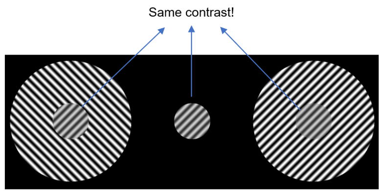
Fig. 1: Surround suppression, caused by a similarly oriented background (on the right side), can decrease the apparent contrast of a visual stimulus. All small (central) circles are identical. Copyright: Sourav/CC-BY-4.0
Surround suppression might have resulted from our brains evolving to process natural stimuli optimally, and might facilitate the segmentation of object boundaries (Angelucci et al., 2017; Coen-Cagli, Dayan, & Schwartz, 2012).
The goal of this master thesis is to uncover neural mechanisms of subjective perception and visual behavior. Thus, you will have the chance to both replicate electrophysiological findings related to surround suppression, and to link it to visual behavioral measures. The work is experimental, involving EEG and behavioral data collection and analysis.
Your profile:
- Strong interest in quantitative experimental research involving participants
- Passion in learning new things
- Ability to work independently
- Nice to have: Basic programming skills in MATLAB/R.
What you can expect:
- A chance to analyze data you will have collected yourself
- Valuable contribution to the scientific community asking for more replication studies, in addition potentially finding novel connections between behavioral and electrophysiological measures
- Experience at a well-established, internationally oriented electrophysiological research laboratory with state of the art equipment
- A balance between supervision and independence
Contact:
Dr. José Ossandón (jose.ossandon"AT"uni-hamburg.de)
References:
Angelucci, A., Bijanzadeh, M., Nurminen, L., Federer, F., Merlin, S., & Bressloff, P. C. (2017). Circuits and mechanisms for surround modulation in visual cortex. Annual Review of Neuroscience, 40(1), 425–451. https://doi.org/10.1146/annurev-neuro-072116-031418
Bair, W., Cavanaugh, J. R., Movshon, J. A., Beest, E. H. van, Grigore, M. E., Levelt, C. N., … Roelfsema, P. R. (2003). Time course and time-distance relationships for surround suppression in macaque V1 neurons. The Journal of Neuroscience : The Official Journal of the Society for Neuroscience, 23(20), 7690–7701. https://doi.org/10.1523/jneurosci.5051-13.2014
Coen-Cagli, R., Dayan, P., & Schwartz, O. (2012). Cortical surround interactions and perceptual salience via natural scene statistics. PLoS Computational Biology, 8(3), e1002405. https://doi.org/10.1371/journal.pcbi.1002405
Vanegas, M. I., Blangero, A., & Kelly, S. P. (2015). Electrophysiological indices of surround suppression in humans. Journal of Neurophysiology, 113(4), 1100–1109. https://doi.org/10.1152/jn.00774.2014
3. EEG Resting State after short-term visual deprivation
We are interested in investigating the excitability of the visual cortex, and how it is adjusted in the short-term via homeostatic plasticity. Previous literature suggests that the effects of visual deprivation on cortical excitability are already apparent after one hour (Boroojerdi, 2000; Fierro et al., 2005; Pitskel et al., 2007). The goal of this experiment is to assess the effects of short-term visual deprivation on cortical excitability, using resting-state electroencephalography (EEG). EEG recordings of resting state activity will be made before and after a short period (1 hour) of total visual deprivation.
The scope of the thesis will include empirical work, that is, EEG data collection, and data analysis.
Contact:
Rashi Pant, M.A. (rashi.pant"AT"uni-hamburg.de)
Dr. José Ossandón (jose.ossandon"AT"uni-hamburg.de)
References:
Boroojerdi B (2000) Enhanced Excitability of the Human Visual Cortex Induced by Short-term Light Deprivation. Cerebral Cortex 10:529–534.
Fierro B, Brighina F, Vitello G, Piazza A, Scalia S, Giglia G, Daniele O, Pascual-Leone A (2005) Modulatory effects of low- and high-frequency repetitive transcranial magnetic stimulation on visual cortex of healthy subjects undergoing light deprivation: rTMS effects on deafferentated visual cortex. The Journal of Physiology 565:659–665.
Pitskel NB, Merabet LB, Ramos-Estebanez C, Kauffman T, Pascual-Leone A (2007) Time-dependent changes in cortical excitability after prolonged visual deprivation. NeuroReport 18:1703–1707.